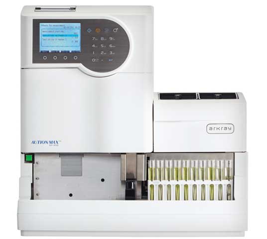
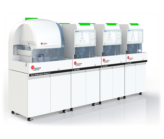
DxU Microscopy Series
Urine Microscopy Analyzer
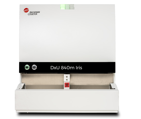
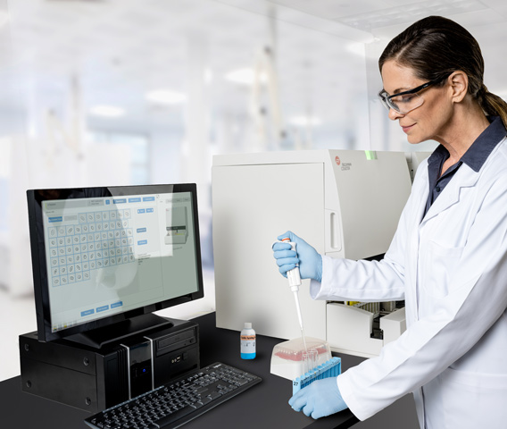
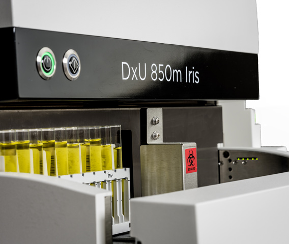
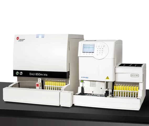
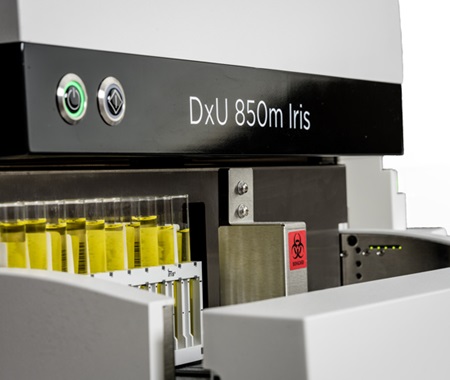
The DxU Iris Microscopy Series automated urine microscopy analyzer includes the DxU 840m Iris, with a throughput of 70 samples per hour or the DxU 850m Iris, with a throughput of 101 samples per hour. The instruments deliver faster turnaround times and accurately standardizes results by reducing manual intervention.
Reduce Manual Reviews
The DxU Iris Workcell streamlines workflow using industry-leading technology to reduce manual review to 4%.1* This is achieved using Proprietary Digital Flow Morphology technology with APR software to capture digital images of particles in the urine or in body fluids. Leveraging this technology reduces total sample processing time up to 78% compared to manual microscopy.2†
Confidence in Results
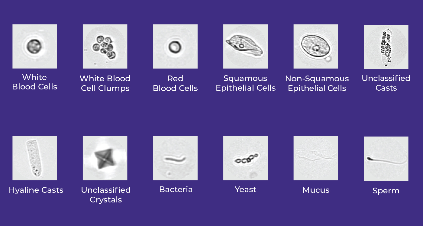
APR Software evaluates the size, shape, contrast and texture of the urine particles and evaluates each digital image to identify and classify the urine particles into one of 12 primary categories. The operator has the option to further subclassify particles into 27 additional categories. This offers easy identification of the urine sediment and quick verification of particle presence reduces the need for microscopy to identify and confirm presence of a category flag or scatterplot. The primary particle categories include:
Auto-release Results
Edit-Free Release technology allows the laboratory to auto-verify true positive urines and true negative urines based on release settings they set up. This feature converts the analyzer into a walkaway unit.
Increase Uptime and Improve Productivity
The iWARE Integrated Urinalysis Software solution, standard on the analyzer, provides onboard validation and result verification into a single step without the need for middleware. The software helps laboratories in the efficient processing of results while reducing time-consuming steps.1‡
Optimize Rinse Routine
The DxU Iris introduces the iQclear, a cleaning aspiration module that helps with additional cleaning of the sample probe.*
Comparison of the DxU Microscopy Series Instruments
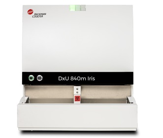
DxU 840m Iris
- 70 samples per hour
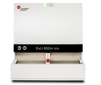
DxU 850m Iris
- 101 samples per hour
| MICROSCOPY | |
Menu/Test parameters |
Urine particles: Additional categories for subclassification:
Body fluids: |
Measurement technology |
Digital Flow Morphology using Auto-Particle Recognition Software |
Sample throughput |
Up to 101 samples per hour |
Sample identification and capacity |
|
Specimen volume |
|
Workstation |
|
Data storage |
Onboard storage of up to 10,000 patient results |
Communication interface |
Bidirectional with host query |
Operating environment |
|
Electrical power requirement |
|
BTU |
1,200 |
Dimensions and Weight |
Depth |
Width |
Height |
Weight |
| Microscopy module | 25.4" (64.5 cm) | 20.9" (53 cm) | 23" (54.4 cm) | 100.0 lbs (46.0 kg) |
| CPU | 10.8" (27.4 cm) | 5.5 in (14.0 cm) | 12.7" (32.3 cm) | 28.0 lbs (13.0 kg) |
| 21.5" Monitor with base | 8.6" (21.8 cm) | 20.4 in (517.4 mm) | 13.8" (35 cm) | 23.8 lbs (10.8 kg) |
| Load/Unload stations | 20.5" (52 cm) | 9.25" (23.5 cm) | 5.7" (14.5 cm) | 11 lbs (5 kg) |
Select Tips & Tools to access helpful documents and information for your instrument.
Step 1: To access content such as job aids, checklists, In-Lab Training Manuals, etc., select the Tips & Tools link in the top left navigation.
Step 2: Select the product name.
Step 3: You will be directed to the product page and access to the product-specific content.
Click here to access Tips & Tools
Automate Body Fluids Analysis
The DxU Micrscopy Series provides a standardized, fully automated method for the analyses of RBC count and nucleated cell count in cerebrospinal, synovial and serous fluids. Digital Flow Morphology technology isolates particles in body fluids to provide immediate and reproducible results that can be verified on the screen.
Learn moreEducational Resources
*The manual review rate was observed at a single site and will vary according to laboratory practices and workflows.
† The reduction in sample processing time was observed at a single site and processing time improvements will vary according to laboratory practices and workflows.
‡This claim is based on a peer-reviewed article.
1. Broadlawns Medical Center. (2019). Manual microscopy divided by on-screen microscopic verification report generated December 20, 2019. Case study published by Beckman Coulter. https://media.beckmancoulter.com/-/media/diagnostics/products/urinalysis/docs/ua-broadlawns-case-study.pdf?_ga=2.105761475.1320714369.1623081858-966792729.1621451088
2. Beaufort Memorial Urinalysis Workflow Case Study, CS-52048.
3. Xuekai L, Weibin C, Xiaolong L, et al. Establishment and validation of auto verification criteria for urine analysis workstation in a multi-center study. Chinese Journal of Laboratory Medicine. December 11, 2020.
The DxUm leverages the same proprietary technology present on the iQ200 series automated urine microscopy analyzers. Results of all studies are transferable.
REMISOL Advance is a trademark or registered trademark of Normand-Info SAS in the United States and other countries. Used under license.
ARKRAY, the stylized logo, and the ARKRAY products mentioned herein are trademarks or registered trademarks of ARKRAY in the United States. 2024-13168
 English


