Understanding Acute Myeloid Leukemia
Acute Myeloid Leukemia (AML) is the most common leukemia among the adult population and accounts for about 80% of all leukemia cases worldwide.2 In 2023, in the United States alone, it is estimated that there are >20,000 new cases of AML,3 with over half of these cases resulting in death. Globally, almost 200,000 cases of AML were reported in 2017, and the numbers are on the rise.2
AML is a serious form of cancer that starts in the bone marrow, but most often moves quickly into the blood. If untreated, it can rapidly spread to other parts of the body including the lymph nodes, liver, spleen, central nervous system, and testicles—leading to death; therefore, it is important to identify and treat it quickly.
The clinical presentation of AML is variable. Severe anemia can manifest in the form of easy fatiguability, shortness of breath, and generalized weakness. Patients may present with recurrent infections, anemia, bruising, bleeding from mucosal sites, and disseminated intravascular coagulation. Patients may also present with signs and symptoms secondary to ineffective erythropoiesis or bone marrow failure. Diagnosis of AML relies on imaging tests and bone marrow tests, but analysis usually begins with blood chemistry and blood work including the CBC-differential (CBC-Diff) test.1
Using Near-native Cellular Characterization to Aid in AML Identification
In this case study, Maite Serrando, M.D., Ph.D., from Laboratori Clinic Territorial IAS-ICS Girona, reviews a patient’s CBC-Diff results from the DxH 900 hematology analyzer and highlights how near-native cellular characterization provided significant data that raised the index of suspicion for AML.
Clinical Laboratory Results:
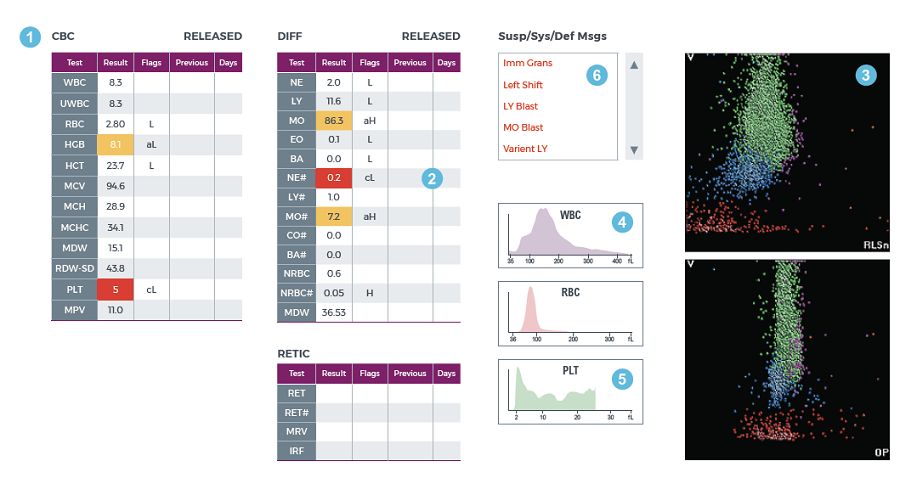
Results of a CBC-Diff from a case-study patient with AML are shown below.
CBC with Differential

- CBC Results: The CBC parameters instrument flags indicate normocytic, normochromic anemia and critically low platelets (thrombocytopenia) without review message
- Automated Differential: The results of the WBC automated differential show monocytosis and critical neutropenia with multiple instrument flags
- Scatter Plot: In this scatter plot there is a single cluster without any boundaries between the Monocyte (green) and Lymphocyte (blue) cell populations (no space between MO and LY), signaling the need for microscopy review
- WBC Histogram: The WBC histogram shows a predominant subpopulation of cells
- PLT Histogram: An abnormal PLT histogram is seen, but the platelet count was reported without review message
- Suspect Message(s): Multiple suspect messages generated by the analyzer suggesting the presence of abnormal cells
The high monocyte count as observed in the CBC-Diff and the lack of space between monocytes and lymphocytes on the scatter plot triggered the need for blood smear review.
Blood Smear Results:
The results in this case study are consistent with those of Acute Myeloid Leukemia. While the blast morphology suggested myeloid lineage, additional diagnostic tests including flow cytometry and genetic testing were used verify AML.
Leveraging the Power of the CBC
In this video, Dr. Serrando discusses the streamlined workflow technology and state-of-the-art pathology-screening capabilities of the DxH 900 Hematology analyzer and how her laboratory applies these data to flag abnormal cells and identify disease. She discusses how test results provide clinically significant data and reviews the CBC-Diff results from a patient with AML.
About Dr. Serrando
Dr. Serrando received her medical degree and laboratory residence from University of Lleida in Catalonia, Spain. Since 2004, Dr. Serrando has worked as a hospital laboratory consultant and currently oversees the Hematology Laboratory Department at Parc Hospitalari Martí i Julià in Salt, Spain. Dr. Serrando has a keen interest in new technologies and has published many papers and presented her work at national and international conferences.
 English


